J Adv Periodontol Implant Dent. 18(3):122-131.
doi: 10.34172/japid.026.3982
Original Article
Salivary antibody responses to Fusobacterium nucleatum and Candida albicans as indicators of periodontitis severity
Muhammad Ihsan Rizal Conceptualization, Data curation, Formal analysis, Funding acquisition, Investigation, Visualization, Writing – original draft, Writing – review & editing, 1, 2, * 
Endang Winiati Bachtiar Conceptualization, Data curation, Methodology, Project administration, Resources, Supervision, Validation, Visualization, Writing – review & editing, 3
Boy Muchlis Bachtiar Funding acquisition, Methodology, Project administration, Resources, Supervision, Validation, Writing – review & editing, 3
Yuniarti Soeroso Data curation, Investigation, Methodology, Project administration, Resources, Supervision, Validation, Writing – review & editing, 4
Fatimah Maria Tadjoedin Data curation, Investigation, Resources, Writing – review & editing, 4
Retno Damajanti Soejoedono Supervision, Writing – review & editing, 5
Ferry Pergamus Gultom Supervision, Writing – review & editing, 3
Natalina Haerani Investigation, Resources, Supervision, Writing – review & editing, 4
Melanie Sadono Djamil Supervision, Writing – review & editing, 1
Boedi Oetomo Roeslan Supervision, Writing – review & editing, 1
Author information:
1Department of Oral Biology, Faculty of Dentistry, Universitas Trisakti, Jakarta, Indonesia
2Center of Molecular Biology Study, Faculty of Dentistry, Universitas Trisakti, Jakarta, Indonesia
3Department of Oral Biology, Faculty of Dentistry, Universitas Indonesia, Jakarta, Indonesia
4Department of Periodontology, Faculty of Dentistry, Universitas Indonesia, Jakarta, Indonesia
5Department of Animal Diseases and Veterinary Health, Medical Microbiology Division, Faculty of Veterinary Medicine, IPB University, Bogor, Indonesia
Abstract
Introduction:
Periodontitis is a chronic inflammatory disease that leads to the destruction of tooth-supporting tissues and remains a major cause of tooth loss worldwide. Its pathogenesis involves complex interactions between microbial pathogens and host immune responses, where salivary immunoglobulins serve as a first line of defense at mucosal surfaces. Identifying pathogen-specific salivary antibody responses that correlate with disease severity may provide noninvasive biomarkers for diagnosis and monitoring. The present work sought to explore how selected microbial pathogens and salivary antibody responses are connected with the severity of periodontitis and clinical periodontal status.
Methods:
A cross-sectional approach was applied. Saliva was obtained from 39 participants, including 31 patients diagnosed with periodontitis and 8 individuals with healthy periodontal tissues. Western blotting and enzyme-linked immunoassays were used to detect both microorganisms and immunoglobulins.
Results:
Patients classified as Stage IV periodontitis showed the highest occurrence of Aggregatibacter actinomycetemcomitans. Elevated IgA antibodies against Fusobacterium nucleatum (P=0.014) and Candida albicans (P=0.009) demonstrated significant associations with disease severity. Further associations were observed: plaque index with IgG to C. albicans; oral hygiene index with IgA to A. actinomycetemcomitans (P=0.008) and C. albicans (P=0.031), and papillary bleeding index with IgA to A. actinomycetemcomitans (P=0.003), F. nucleatum (P=0.002), and C. albicans (P=0.008).
Conclusion:
Salivary IgA and IgG responses to Fusobacterium nucleatum and Candida albicans exhibited significant stage-related associations with periodontitis severity, supporting their potential role as complementary immunological indicators.
Graphical Abstract
Keywords: Humoral immune response, Keystone pathogen, Periodontitis, Polymicrobial, Saliva
Copyright and License Information
© 2026 The Author(s).
This is an open access article distributed under the terms of the Creative Commons Attribution License (
http://creativecommons.org/licenses/by/4.0/), which permits unrestricted use, distribution, and reproduction in any medium, provided the original work is properly cited.
Funding Statement
This research was funded by Universitas Trisakti Grant no: 82/FKG/4/09/2018.
Introduction
The condition known as periodontitis involves progressive destruction of the structures that support teeth, such as the periodontal ligament, gingival tissues, cementum, and alveolar bone.1,2 Data from the World Health Organization highlight this disorder as a major contributor to tooth loss, affecting roughly one-fifth of the adult population globally.3 Other epidemiological evidence has suggested a wider prevalence, estimated between 20% and 50% worldwide.4 The Global Burden of Disease Study (2016) placed severe forms of periodontal disease as the sixth most widespread health problem globally.5 As scientific understanding of its pathogenesis has deepened, and definitions and classifications of periodontitis have been repeatedly updated. The most recent revision, issued in 2017, stratifies cases into four stages—ranging from Stage I through Stage IV—according to both clinical severity and treatment considerations.6
The pathogenesis of periodontitis has been interpreted through multiple conceptual models, including nonspecific and specific plaque hypotheses, the ecological plaque perspective, the keystone pathogen model, and most recently, the polymicrobial synergy and dysbiosis (PSD) framework. The keystone pathogen concept emphasizes that certain microbes, even when present at low abundance, can reprogram the oral microbiota toward dysbiosis and undermine host defenses, in part by deriving sustenance from epithelial cells.7 Central to this idea is the ability of such organisms to interfere with immune function, especially with neutrophils positioned between dental biofilms and the epithelial layer. Porphyromonas gingivalis has long been regarded as the archetypal keystone pathogen in periodontitis.8 Later, Lamont et al.9 refined this concept into the PSD hypothesis.9 Findings from animal experiments confirmed that mixed infections by periopathogens exhibit markedly stronger pathogenicity compared with single-species infections. This increased virulence likely reflects the requirement of multiple bacterial factors—including adhesins, receptors, proteases, and proinflammatory surface molecules—that are absent in single keystone strains. Acting together, these elements constitute a collective virulence trait that promotes persistent dysbiosis, triggers inflammatory responses, and ultimately contributes to periodontal tissue breakdown.10
In an investigation of salivary proteomics in periodontitis, six proteins were found to be elevated in individuals with aggressive disease compared with healthy controls: serum albumin, immunoglobulin A (IgA), immunoglobulin G (IgG), vitamin D-binding protein, salivary α-amylase, and zinc-α2 glycoprotein.11 IgA contributes significantly to defense against bacterial and viral pathogens, while IgG, a serum-derived antibody, specifically targets microorganisms implicated in periodontal disease. Both immunoglobulins arise from the adaptive immune response, which is orchestrated by activated B and T lymphocytes.12 These lymphocytes express receptors that recognize antigens—unique microbial or host-derived molecules—thereby enabling the immune system to respond with remarkable specificity.13
Within salivary glands, plasma cells produce IgA, making it the most prevalent antibody in saliva.14,15 IgG, in contrast, is mainly synthesized within gingival tissues; during inflammation, B lymphocytes differentiate into plasma cells and release IgG locally. Circulating antibodies can also access the oral cavity through gingival crevicular fluid. In addition to immunoglobulins, saliva contains multiple innate antimicrobial factors secreted by salivary glands, epithelial cells, and neutrophils. These substances restrict or eliminate bacterial, fungal, and viral growth.13
Despite growing interest in salivary biomarkers in periodontitis, most previous investigations have focused on single microorganisms or isolated host markers, often without integrating microbial detection with humoral immune responses across clinically stratified disease stages. For this purpose, the following microorganisms were chosen as representative antigens known to elicit adaptive immune reactions: Aggregatibacter actinomycetemcomitans, Treponema denticola, Porphyromonas gingivalis, Fusobacterium nucleatum, and Candida albicans. Furthermore, few studies have evaluated salivary antibody profiles in the context of the 2017 AAP/EFP classification system, which provides a more refined staging framework for disease severity. As a result, the relationship between polymicrobial presence, salivary immunoglobulin responses, and stage-specific clinical parameters remains insufficiently characterized. Therefore, this study aimed to evaluate salivary humoral immunologic markers and microbial presence across severity stages of periodontitis defined by the 2017 AAP/EFP classification.
Methods
This investigation followed an observational, cross-sectional design. It was carried out at three locations: the Dental Hospital of Universitas Indonesia, the Biomolecular Laboratory of the Faculty of Dentistry at Universitas Indonesia, and the BioCORE Laboratory of the Faculty of Dentistry at Universitas Trisakti. A priori sample size estimation was conducted using the Lemeshow formula for unpaired numerical analysis. With a significance level set at 5% (α = 0.05) and statistical power of 80%, and based on the anticipated standard deviation and expected mean difference between groups, the minimum sample size required was five subjects per group. Eligible participants were men and women aged 18–60 years, who visited the Universitas Indonesia Dental Hospital. The subjects were categorized as periodontitis patients (Stages I–IV) or individuals with clinically healthy periodontium. Only individuals willing to participate and provide written informed consent were enrolled. Participants were excluded if they had a history of systemic diseases, including hypertension, cardiovascular disease, kidney disease, or hematological disorders; had consumed systemic medications within the past three months; were pregnant or breastfeeding; or had received dental treatment within the previous three months.
This study was approved by the Ethics Committee of the Faculty of Dentistry, Universitas Indonesia (Approval No. 17/Ethical Approval/FKGUI/III/2019; Protocol No. 070210219). All the participants provided written informed consent prior to enrolment. Demographic data were recorded, and each subject underwent a general health evaluation and a comprehensive periodontal examination. The following parameters were documented: plaque index, calculus index, papillary bleeding index, oral hygiene index, pocket depth, gingival recession, clinical attachment loss, and tooth count. Classification of disease severity followed the 2017 framework jointly proposed by the American Academy of Periodontology (AAP) and the European Federation of Periodontology (EFP).
Salivary Protein Preparation
Unstimulated morning salivary samples (volume: < 2 mL) were collected into 15-mL tubes. To minimize protein degradation, samples were immediately placed on ice, and 50 µL of a protease inhibitor cocktail was added. Homogenization was performed by centrifugation (1300 × g, 5 min, 4°C). Supernatant aliquots (150 µL) were transferred into new microtubes and subjected to protein extraction using GenezolTM reagent (GeneAid, Taiwan). Salivary protein samples were stored at -20°C prior to analysis to preserve protein integrity. Protein concentrations were then determined with the Bradford assay (Bio-Rad, USA), according to the manufacturer’s protocol.
Western Blotting
Western blotting was performed under denaturing and reducing conditions using SDS-PAGE, followed by semi-dry transfer to PVDF membranes and indirect HRP-based chromogenic detection. Rabbit polyclonal antibodies against Aggregatibacter actinomycetemcomitans, Treponema denticola, Porphyromonas gingivalis, Fusobacterium nucleatum, and Candida albicans were generated at the Faculty of Veterinary Medicine, IPB University. These antibodies had been purified previously using a high-affinity purification kit (GenScript, USA) and validated for specificity through dot blotting. Protein samples ( ≤ 3 μg) were mixed with buffer (Bio-Rad, USA) and β-mercaptoethanol (Sigma-Aldrich, USA) at a 19:1 ratio and then diluted 1:1 with sample buffer to a final volume of 20 μL. Following denaturation at 100°C for 5 min, 20 μL of each preparation was loaded into polyacrylamide gels along with pre-stained molecular weight standards (Precision Plus ProteinTM Dual Color Standards, Bio-Rad, USA). Electrophoresis was conducted at 90 V for 15 min and then at 125 V for 1 h. Proteins were transferred to PVDF membranes using a Trans-Blot Turbo semi-dry system (Bio-Rad, USA). Goat anti-rabbit IgG conjugated to HRP (1:1000, 5% non-fat milk; Thermo Fisher Scientific, USA) served as the secondary antibody. Protein visualization was achieved using the 1-StepTM Chloronaphthol Substrate Solution (Thermo Fisher Scientific, USA), and band intensities were quantified with ImageJ software.
ELISA for Salivary IgA and IgG
Indirect enzyme-linked immunoassays (ELISA) were performed to assess IgA and IgG reactivity against the selected pathogens. Wells were first coated with 100 μL of carbonate buffer (3.7 g of sodium bicarbonate and 0.64 g of sodium carbonate in 1 L of distilled water). Pathogens were added in 50-μL aliquots at the following concentrations: A. actinomycetemcomitans, 5.4 × 10⁷ CFU/mL; T. denticola (ATCC® 35405), 2 × 10⁸ CFU/mL; P. gingivalis (ATCC® 33277), 1.3 × 10⁷ CFU/mL; F. nucleatum (ATCC® 25586), 3.89 × 10⁸ CFU/mL; and C. albicans (ATCC® 10231), 5.9 × 10⁷ CFU/mL. Plates were incubated overnight at 4°C with 5% CO₂. After incubation, the wells were emptied and washed three times with 200 μL of TBS-T buffer. Blocking was performed with 150 μL of 5% non-fat milk for 1 h at 37°C and 5% CO₂. After washing, 50 μL of saliva from each participant was added and incubated for 1 h under the same conditions. The plates were washed again, and 100 μL of HRP-conjugated secondary antibodies (anti-human IgA and anti-human IgG, 1:2000 in 5% non-fat milk; Thermo Fisher Scientific, USA) was added. Absorbance was measured at 450 nm using an iMarkTM Microplate Reader (Bio-Rad, USA). Cut-off values for IgA and IgG responses were determined using receiver operating characteristic (ROC) curve analysis.
Statistical Analysis
Data were analyzed using SPSS® 23. Descriptive statistics summarized demographic and clinical characteristics. Normality of continuous variables, including clinical indices, salivary protein concentration, and antibody optical density values, was assessed using the Shapiro–Wilk test. Inter-group comparisons across periodontitis stages were performed using one-way ANOVA with post hoc Bonferroni correction for normally distributed data and the Kruskal–Wallis test followed by the Mann–Whitney U test for non-normally distributed data. Associations between categorical variables, including antibody response categories and clinical index classifications, were evaluated using the chi-squared test, with odds ratios (OR) and 95% confidence intervals (CI) calculated where appropriate. Spearman’s rank correlation was specifically applied to assess the relationship between the presence of Aggregatibacter actinomycetemcomitans antigen and salivary IgA and IgG responses in Stage III periodontitis subjects. Statistical significance was set at P < 0.05.
Results
Thirty-nine individuals were included in this study, 12 of whom were men (30.7%) and 27 were women (69.2%). Using the 2017 AAP/EFP classification, 31 participants were diagnosed with periodontitis at different stages, while eight were periodontally healthy. As detailed in Table 1, subjects with more advanced stages tended to be older, with mean age rising from 25.1 ± 2.9 years in the healthy group to 51.0 ± 6.1 years in Stage IV. Indices reflecting oral health status showed a clear gradient: plaque index, oral hygiene index, and papillary bleeding index increased progressively with disease severity. For instance, the plaque index rose from 0.45 ± 0.13 in healthy subjects to 1.58 ± 0.30 in Stage IV. By contrast, salivary protein concentrations remained relatively stable, ranging from 147.38 ± 4.21 to 158.38 ± 12.10 µg/mL, suggesting that total protein content in saliva was not influenced by disease stage.
Table 1.
Characteristics of participants stratified by periodontitis severity. Variables include gender distribution, mean age, plaque index, oral hygiene index, papillary bleeding index, and salivary protein concentration
|
Periodontitis Stage (n)
|
Gender
(M/F)
|
Mean Age
±SD
|
Mean PI
±SD
|
Mean OHI
±SD
|
Mean PBI
±SD
|
Salivary protein
±SD (µg/mL)
|
| Healthy (8) |
1/7 |
25.1 ± 2.9 |
0.45 ± 0.13 |
0.25 ± 0.21 |
0.10 ± 0.09 |
147.38 ± 4.21 |
| Stage I (5) |
2/3 |
41.8 ± 5.3 |
0.69 ± 0.25 |
1.05 ± 0.47 |
0.67 ± 0.45 |
153.24 ± 4.55 |
| Stage II (8) |
3/5 |
45.1 ± 7.3 |
0.82 ± 0.27 |
1.33 ± 0.45 |
0.92 ± 0.59 |
156.37 ± 11.34 |
| Stage III (11) |
4/7 |
45.9 ± 8.3 |
1.27 ± 0.35 |
2.68 ± 0.57 |
1.67 ± 0.54 |
158.38 ± 12.10 |
| Stage IV (7) |
2/5 |
51.0 ± 6.1 |
1.58 ± 0.30 |
3.48 ± 0.54 |
2.59 ± 0.25 |
155.57 ± 7.03 |
SD: standard deviation; M: male; F: female; PI: plaque index; OHI: oral hygiene index; PBI: papilla bleeding index
Detection of A. actinomycetemcomitans
Western blot analysis identified A. actinomycetemcomitans antigen in the saliva of all study participants. Quantification of band density using ImageJ software revealed the highest median values among Stage IV cases (8096.35; range: 1134.08–14587.27), compared with 7594.34 in the healthy group. However, this apparent trend was not statistically significant (Kruskal–Wallis, P = 0.453) (Figure 1), indicating that although A. actinomycetemcomitans was present across groups, its abundance alone may not directly reflect disease severity.
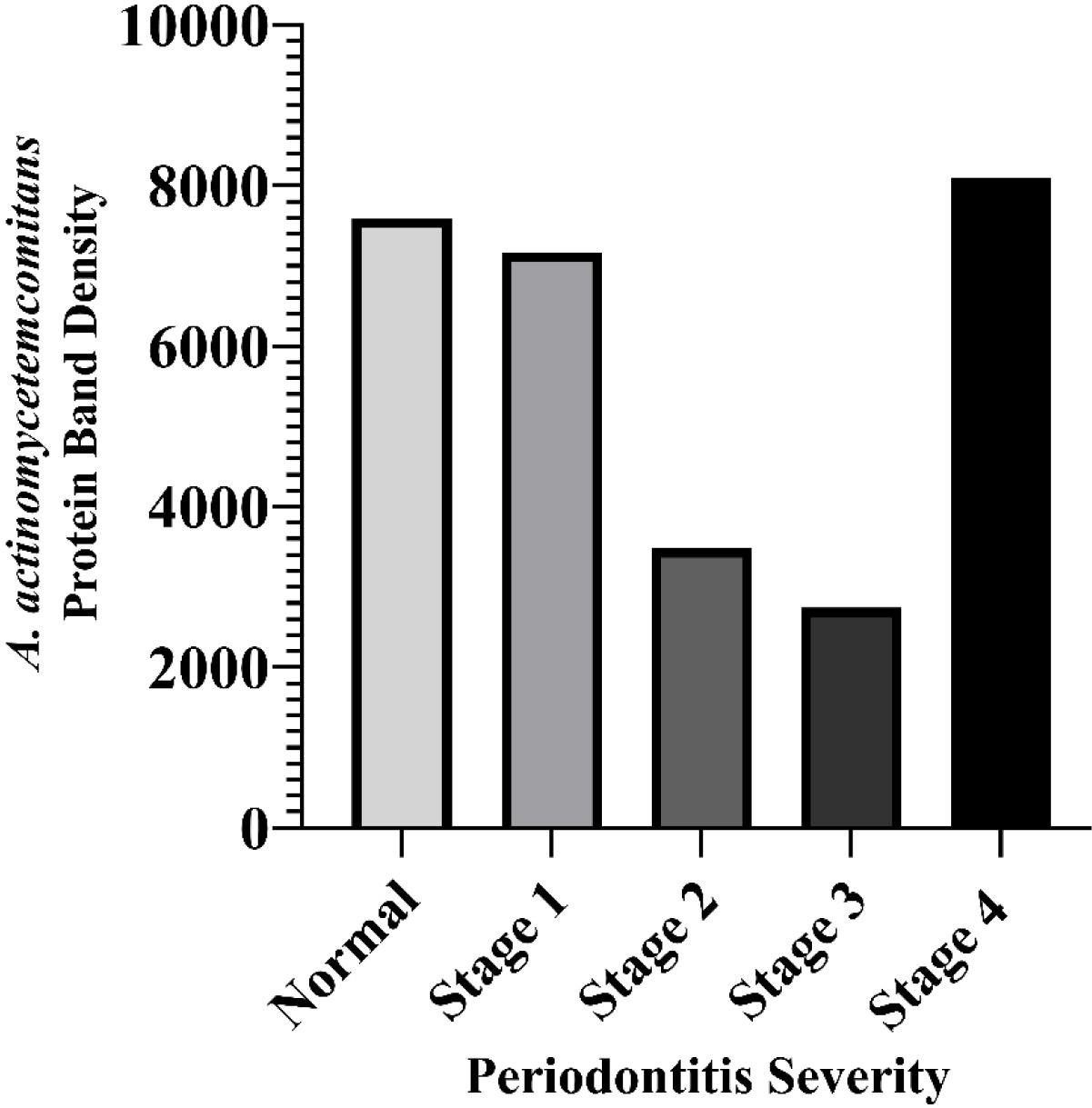
Figure 1.
ImageJ analysis of protein band density in the western blot assay of Aggregatibacter actinomycetemcomitans antigen in saliva based on periodontitis severity. Median (min-max) normal: 7594.34 (1867.27-11343.25); Stage I: 7170.35 (1417.93-14,639.41); Stage II: 3482.51 (600.33-20.242.59); Stage III: 2742.23 (1009.98-10925.25); Stage IV: 8096.35 (1134.08-14,587.27). Kruskal-Wallis test P = 0.453
.
ImageJ analysis of protein band density in the western blot assay of Aggregatibacter actinomycetemcomitans antigen in saliva based on periodontitis severity. Median (min-max) normal: 7594.34 (1867.27-11343.25); Stage I: 7170.35 (1417.93-14,639.41); Stage II: 3482.51 (600.33-20.242.59); Stage III: 2742.23 (1009.98-10925.25); Stage IV: 8096.35 (1134.08-14,587.27). Kruskal-Wallis test P = 0.453
Salivary IgA Responses
The analysis of IgA responses demonstrated stage-dependent variability. Significant associations were observed for F. nucleatum (P = 0.014) and C. albicans (P = 0.009), with antibody levels peaking in Stage IV subjects (Figure 2). Median IgA optical density values for F. nucleatum ranged from 1.15 in Stage I to 3.44 in Stage IV, while those for C. albicans increased from 0.74 in Stage I to 2.46 in Stage IV. In contrast, IgA responses to A. actinomycetemcomitans, T. denticola, and P. gingivalis did not differ significantly between the groups, suggesting that the IgA response may be more selectively amplified against certain pathogens.
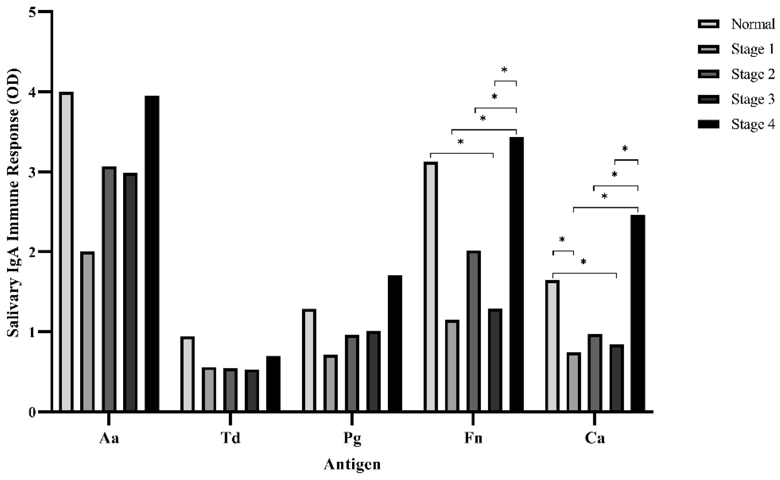
Figure 2.
Median values of salivary IgA immune response to periodontal bacterial antigens and Candida albicans based on periodontitis severity. Kruskal‒Wallis test and post hoc Mann‒Whitney U test (* = significantly different; P < 0.05).
Aa: Aggregatibacter actinomycetemcomitans; Td: Treponema denticola; Pg: Porphyromonas gingivalis; Fn: Fusobacterium nucleatum; Ca: Candida albicans; OD: optical density
.
Median values of salivary IgA immune response to periodontal bacterial antigens and Candida albicans based on periodontitis severity. Kruskal‒Wallis test and post hoc Mann‒Whitney U test (* = significantly different; P < 0.05).
Aa: Aggregatibacter actinomycetemcomitans; Td: Treponema denticola; Pg: Porphyromonas gingivalis; Fn: Fusobacterium nucleatum; Ca: Candida albicans; OD: optical density
Salivary IgG Responses
A similar pattern was found for IgG. Responses to F. nucleatum (P = 0.011) and C. albicans (P = 0.015) increased significantly with disease severity (Figure 3). In Stage IV patients, IgG reactivity reached median optical density values of 1.29 for F. nucleatum and 0.51 for C. albicans, compared with 1.14 and 0.20, respectively, in healthy controls. No significant stage-related differences were observed in IgG responses to A. actinomycetemcomitans, T. denticola, or P. gingivalis. These findings suggest that both IgA and IgG immune responses to F. nucleatum and C. albicans are closely linked to disease progression.
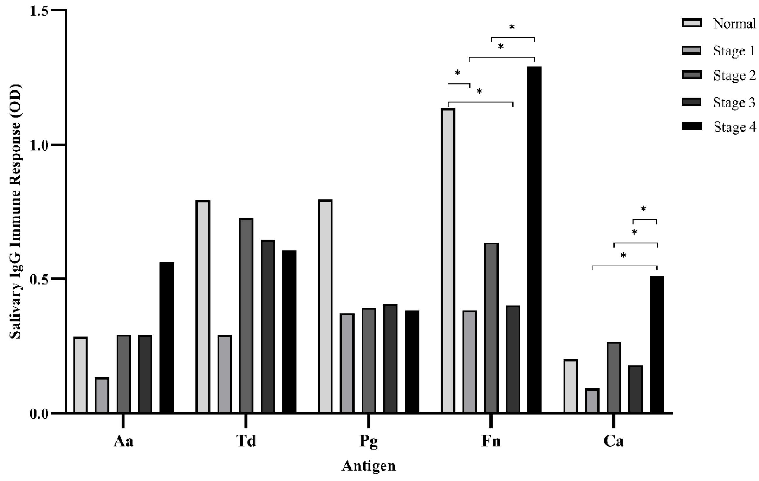
Figure 3.
Median values of salivary IgG immune response to periodontal bacterial antigens and Candida albicans based on periodontitis severity. Kruskal–Wallis test and post hoc Mann-Whitney U test (* = significantly different; P < 0.05).
Aa: Aggregatibacter actinomycetemcomitans; Td: Treponema denticola; Pg: Porphyromonas gingivalis; Fn: Fusobacterium nucleatum; Ca: Candida albicans; OD: optical density
.
Median values of salivary IgG immune response to periodontal bacterial antigens and Candida albicans based on periodontitis severity. Kruskal–Wallis test and post hoc Mann-Whitney U test (* = significantly different; P < 0.05).
Aa: Aggregatibacter actinomycetemcomitans; Td: Treponema denticola; Pg: Porphyromonas gingivalis; Fn: Fusobacterium nucleatum; Ca: Candida albicans; OD: optical density
Correlation of A. actinomycetemcomitans with Antibody Responses
To explore whether bacterial presence was related to salivary antibody responses, Spearman’s test was performed. In Stage III subjects, the presence of A. actinomycetemcomitans showed strong positive correlations with both IgA and IgG responses (Figure 4). This finding suggests that the bacterium may stimulate local antibody production, particularly in intermediate stages of disease, when teeth are still present and immune interaction is active.
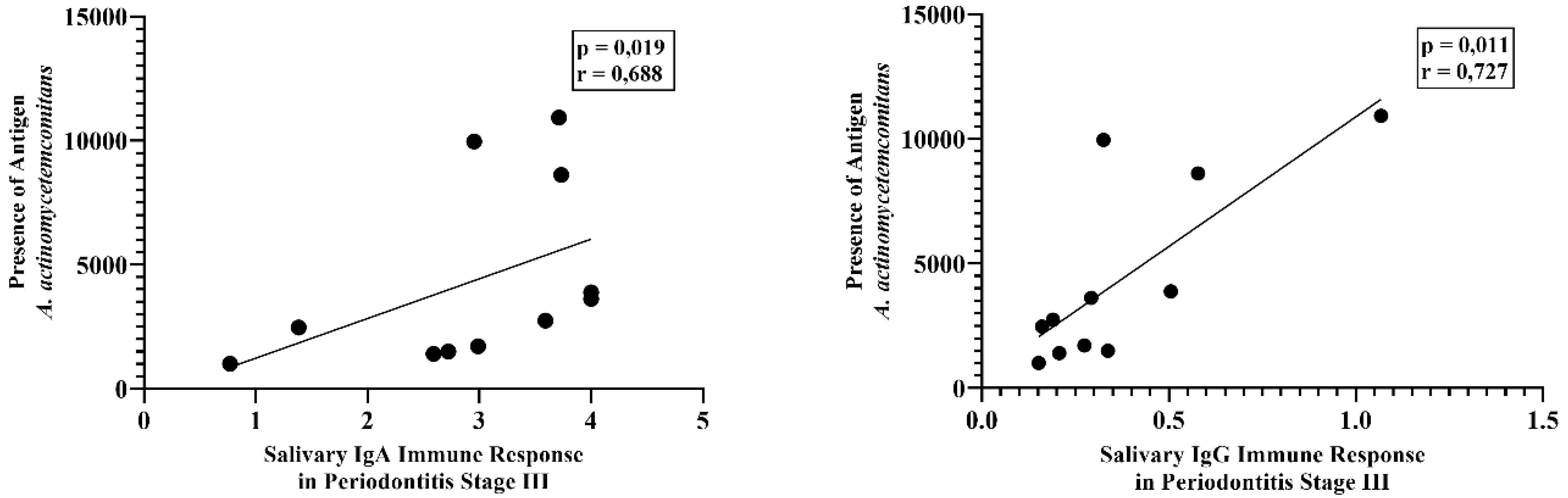
Figure 4.
Relationship between the presence of Aggregatibacter actinomycetemcomitans antigen with IgA (left) and IgG (right) salivary immune responses in periodontitis stage III subjects.
Description: Spearman’s test; IgA: immunoglobulin A; IgG: immunoglobulin G
.
Relationship between the presence of Aggregatibacter actinomycetemcomitans antigen with IgA (left) and IgG (right) salivary immune responses in periodontitis stage III subjects.
Description: Spearman’s test; IgA: immunoglobulin A; IgG: immunoglobulin G
Clinical Indices and Disease Severity
Comparison of clinical indices confirmed significant differences across disease stages. Plaque index, oral hygiene index, and papillary bleeding index were all strongly associated with severity (ANOVA/Kruskal–Wallis, all P < 0.001) (Figure 5). These indices therefore reflect not only disease stage but also the host’s inflammatory status.
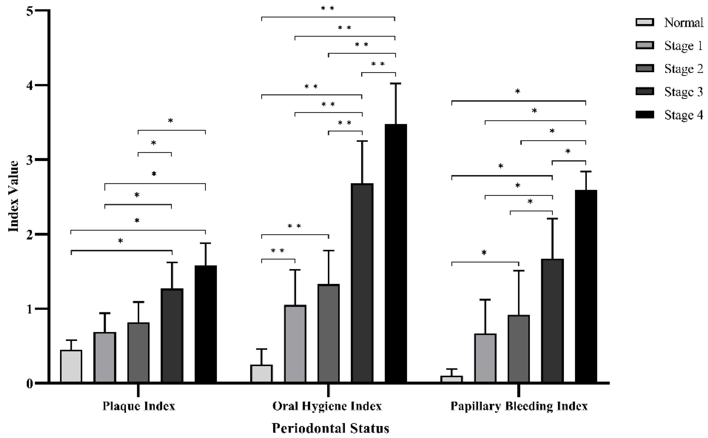
Figure 5.
Mean values of periodontal status (plaque index, oral hygiene index, and papillary bleeding index) in periodontitis severity. ANOVA test and post hoc Bonferroni test (* = significantly different; P < 0.05). Kruskal–Wallis Test and post hoc Mann‒Whitney U Test (** = significantly different; P < 0.05)
.
Mean values of periodontal status (plaque index, oral hygiene index, and papillary bleeding index) in periodontitis severity. ANOVA test and post hoc Bonferroni test (* = significantly different; P < 0.05). Kruskal–Wallis Test and post hoc Mann‒Whitney U Test (** = significantly different; P < 0.05)
Clinical Indices and Immune Responses
Further analysis was performed to assess whether clinical indices were linked to salivary immune responses. As presented in Table 2, plaque index was significantly correlated with IgG reactivity to C. albicans (P = 0.041, OR = 5.333, 95% CI: 1.175–24.213), suggesting that increased plaque accumulation may enhance the humoral response to this fungal pathogen. In contrast, no statistically significant differences were found for other pathogens in the IgG analysis, nor were any significant associations observed in the IgA responses (P > 0.05).
Table 2.
Correlation between plaque category and salivary IgA/IgG responses to selected periodontal pathogens
|
Antibody
|
Pathogens
|
High response
n (%)
|
Low response
n (%)
|
Plaque
category
|
P
-value
|
OR
|
95% CI
|
| IgA |
A. actinomycetemcomitans
|
6 (54.5)/7 (25.0) |
5 (45.5)/21 (75.0) |
Thin vs. Moderate |
0.131 |
0.278 |
0.064–1.200 |
|
T. denticola
|
9 (45.0)/4 (21.1) |
11 (55.0)/15 (78.9) |
Thin vs. Moderate |
0.176 |
0.326 |
0.079–1.337 |
|
P. gingivalis
|
2 (20.0)/11 (37.9) |
8 (80.0)/18 (62.1) |
Thin vs. Moderate |
0.445 |
2.444 |
0.437–13.672 |
|
F. nucleatum
|
9 (45.0)/4 (21.1) |
11 (55.0)/15 (78.9) |
Thin vs. Moderate |
0.176 |
0.326 |
0.079–1.337 |
|
C. albicans
|
8 (44.4)/5 (23.8) |
10 (55.6)/16 (76.2) |
Thin vs. Moderate |
0.196 |
0.391 |
0.099–1.535 |
| IgG |
A. actinomycetemcomitans
|
4 (19.0)/9 (50.0) |
17 (81.0)/9 (50.0) |
Thin vs. Moderate |
0.087 |
4.250 |
1.019–17.729 |
|
T. denticola
|
7 (26.9)/6 (46.2) |
19 (73.1)/7 (53.8) |
Thin vs. Moderate |
0.290 |
2.327 |
0.578–9.367 |
|
P. gingivalis
|
8 (50.0)/5 (21.7) |
8 (50.0)/18 (78.3) |
Thin vs. Moderate |
0.090 |
0.278 |
0.069–1.119 |
|
F. nucleatum
|
9 (42.9)/4 (22.2) |
12 (57.1)/14 (77.8) |
Thin vs. Moderate |
0.307 |
0.381 |
0.093–1.557 |
|
C. albicans
|
3 (15.8)/10 (50.0) |
16 (84.2)/10 (50.0) |
Thin vs. Moderate |
0.041
|
5.333 |
1.175–24.213 |
OR: odds ratio; CI: confidence interval; chi-squared test significant values (P < 0.05) are shown in bold
Oral hygiene index demonstrated significant associations with IgA responses to A. actinomycetemcomitans (P = 0.008, OR = 0.112, 95% CI: 0.022–0.567) and C. albicans (P = 0.031, OR = 0.153, 95% CI: 0.027–0.854) (Table 3). Poorer oral hygiene was thus linked to stronger local antibody responses against these organisms.
Table 3.
Correlation between oral hygiene category and salivary IgA/IgG responses to selected periodontal pathogens
|
Antibody
|
Pathogens
|
High response
n (%)
|
Low response
n (%)
|
Oral hygiene
category
|
P-value
|
OR
|
95% CI
|
| IgA |
A. actinomycetemcomitans
|
7 (53.8)/3 (11.5) |
6 (46.2)/23 (88.5) |
Good vs. Fair–Poor |
0.008
|
0.112 |
0.022–0.567 |
|
T. denticola
|
8 (40.0)/2 (10.5) |
12 (60.0)/17 (89.5) |
Good vs. Fair–Poor |
0.065 |
0.176 |
0.032–0.982 |
|
P. gingivalis
|
9 (37.5)/1 (6.7) |
15 (62.5)/14 (93.3) |
Good vs. Fair–Poor |
0.057 |
0.119 |
0.013–1.064 |
|
F. nucleatum
|
8 (40.0)/2 (10.5) |
12 (60.0)/17 (89.5) |
Good vs. Fair–Poor |
0.065 |
0.176 |
0.032–0.982 |
|
C. albicans
|
8 (42.1)/2 (10.0) |
11 (57.9)/18 (90.0) |
Good vs. Fair–Poor |
0.031
|
0.153 |
0.027–0.854 |
| IgG |
A. actinomycetemcomitans
|
3 (17.6)/7 (31.8) |
14 (82.4)/15 (68.2) |
Good vs. Fair–Poor |
0.464 |
2.187 |
0.469–10.119 |
|
T. denticola
|
7 (38.9)/3 (14.3) |
11 (61.1)/18 (85.7) |
Good vs. Fair–Poor |
0.141 |
0.262 |
0.056–1.230 |
|
P. gingivalis
|
6 (37.5)/4 (17.4) |
10 (62.5)/19 (82.6) |
Good vs. Fair–Poor |
0.264 |
0.351 |
0.080–1.540 |
|
F. nucleatum
|
8 (40.0)/2 (10.5) |
12 (60.0)/17 (89.5) |
Good vs. Fair–Poor |
0.065 |
0.176 |
0.032–0.982 |
|
C. albicans
|
3 (16.7)/7 (33.3) |
15 (83.3)/14 (66.7) |
Good vs. Fair–Poor |
0.290 |
2.500 |
0.538–11.617 |
*OR: odds ratio; CI: confidence interval; chi-squared test significant values (P < 0.05) are shown in bold
Papillary bleeding index showed even broader associations, being significantly related to IgA responses against A. actinomycetemcomitans (P = 0.003, OR = 0.071), F. nucleatum (P = 0.020, OR = 0.083), and C. albicans (P = 0.008, OR = 0.072) (Table 4). In contrast, IgG responses did not show significant correlations with papillary bleeding. This indicates that IgA, as the predominant mucosal antibody, may play a more direct role in the local inflammatory processes associated with gingival bleeding. For all analyses, odds ratios below 1.0 reflect inverse associations based on the selected reference categories and should not be interpreted as protective effects.
Table 4.
Correlation between papillary bleeding category and salivary IgA/IgG responses to selected periodontal pathogens
|
Antibody
|
Pathogens
|
High response
n (%)
|
Low response
n (%)
|
Papillary bleeding
category
|
P-value
|
OR
|
95% CI
|
| IgA |
A. actinomycetemcomitans
|
7 (53.8)/2 (7.7) |
6 (46.2)/24 (92.3) |
Good vs. Fair–Poor |
0.003
|
0.071 |
0.012–0.436 |
|
T. denticola
|
7 (38.9)/2 (9.5) |
11 (61.1)/19 (90.5) |
Good vs. Fair–Poor |
0.055 |
0.165 |
0.029–0.941 |
|
P. gingivalis
|
8 (34.8)/1 (6.3) |
15 (65.2)/15 (93.8) |
Good vs. Fair–Poor |
0.056 |
0.125 |
0.014–1.127 |
|
F. nucleatum
|
8 (40.0)/1 (5.3) |
12 (60.0)/18 (94.7) |
Good vs. Fair–Poor |
0.020
|
0.083 |
0.009–0.775 |
|
C. albicans
|
8 (42.1)/1 (5.0) |
11 (57.9)/19 (95.0) |
Good vs. Fair–Poor |
0.008
|
0.072 |
0.008–0.658 |
| IgG |
A. actinomycetemcomitans
|
6 (26.1)/2 (13.3) |
17 (73.9)/13 (86.7) |
Good vs. Fair–Poor |
0.440 |
0.436 |
0.075–2.523 |
|
T. denticola
|
7 (38.9)/2 (9.5) |
11 (61.1)/19 (90.5) |
Good vs. Fair–Poor |
0.055 |
0.165 |
0.029–0.941 |
|
P. gingivalis
|
4 (40.0)/5 (17.2) |
6 (60.0)/24 (82.8) |
Good vs. Fair–Poor |
0.197 |
0.313 |
0.064–1.533 |
|
F. nucleatum
|
3 (37.5)/6 (19.4) |
5 (62.5)/25 (80.6) |
Good vs. Fair–Poor |
0.355 |
0.400 |
0.074–2.159 |
|
C. albicans
|
3 (42.9)/6 (18.8) |
4 (57.1)/26 (81.3) |
Good vs. Fair–Poor |
0.319 |
0.308 |
0.054–1.754 |
OR: odds ratio; CI: confidence interval; chi-squared test significant values (P < 0.05) are shown in bold
Discussion
The present findings revealed that Aggregatibacter actinomycetemcomitans was most abundant in subjects with Stage IV periodontitis. This aligns with earlier studies demonstrating that this bacterium produces quorum-sensing autoinducer-2 molecules in vitro, which interfere with Candida albicans hyphal formation and biofilm development.16,17 In addition, significant associations were observed between periodontitis severity and salivary IgA and IgG directed against Fusobacterium nucleatum and C. albicans. F. nucleatum, a representative of the orange complex, is an opportunistic pathogen well known for its adhesive capacity. By binding to various bacterial species, it acts as a bridge between commensals and pathogenic organisms within dental biofilms.18
The role of orange complex bacteria is crucial for sustaining the red complex, as the latter depends on their presence for survival in the oral cavity.19 Adhesins such as FadA have intensified interest in the pathogenic role of F. nucleatum.20 Its involvement has been linked not only to periodontitis but also to multiple systemic disorders, including cardiovascular disease, adverse pregnancy outcomes, gastrointestinal infections, rheumatoid arthritis, diabetes mellitus, and gastric cancer, where it has even been suggested as a potential diagnostic marker.21 FadA facilitates binding to endothelial cells and promotes both pericellular and intracellular invasion, while also increasing endothelial permeability—mechanisms that may explain its systemic dissemination.22
Although F. nucleatum is often susceptible to cytokine activity and phagocytosis, under certain conditions, it can stimulate inflammation through elevated production of proinflammatory cytokines and metalloproteinases. Molecules such as IL-8, MMP-9, and MMP-13 are particularly important in regulating migration and survival of infected epithelial cells.23 The bacterium can further modulate immunity by inducing apoptosis in circulating polymorphonuclear and mononuclear cells and impairing B- and T-lymphocyte function.24 Neutrophil function is also altered, with enhanced phagocytosis reported.25 Moreover, its serine proteases are able to degrade the IgA α-chain, allowing immune evasion.26
Interestingly, the IgA and IgG responses to F. nucleatum in Stage IV cases were not markedly different from those in healthy controls. This parallels the work of Papapanou et al.,27 who observed that serum IgG titers against periodontal bacteria remained stable over extended periods and correlated with disease progression, though titers may temporarily decline following therapy. Host susceptibility, however, appears to be a critical factor in determining whether microbial imbalance progresses to destructive inflammation. Individuals able to tolerate the shift from symbiosis to dysbiosis may harbor dysbiotic microbiota without developing irreversible periodontal breakdown.28
In the oral cavity, C. albicans colonizes mucosal surfaces, saliva, and periodontal pockets, suggesting a role in disease development.29 Its adaptability, including morphological switching between yeast and hyphal forms, contributes to survival and pathogenicity.30 Evidence from Canabarro et al.31 further supports its role in driving disease severity.31 In the present study, IgA and IgG reactivity to C. albicans increased with severity. Notably, F. nucleatum demonstrates strong adherence to C. albicans, mediated by carbohydrate–protein interactions between the two species.32,33
Innate defenses usually prevent fungal overgrowth, including salivary flow, antimicrobial peptides like histatins, and competition with bacteria for nutrients. Activation of complement through alternative pathways, opsonization, and subsequent recognition by immune cells constitute another protective mechanism. Recognition of C. albicans pathogen-associated molecular patterns (PAMPs) by antigen-presenting cells via pattern recognition receptors, particularly TLR2 and TLR4, initiates both innate and adaptive immune cascades.34
For Porphyromonas gingivalis, salivary IgA and IgG responses showed no association with severity. This observation supports the keystone pathogen hypothesis, which proposes that P. gingivalis manipulates the immune system to promote dysbiosis.7 Animal experiments confirm that low colonization levels of P. gingivalis can trigger inflammation and bone resorption only when accompanied by other microbes, as it fails to cause disease alone in germ-free rats.35 Virulence requires interaction with host immune receptors; strains lacking specific receptors cannot induce dysbiosis in mouse models.7,36 Gingipains, the Arg-specific cysteine proteases of P. gingivalis, activate complement C5a receptors and interact with TLR2, producing inflammatory responses while simultaneously impairing leukocyte bactericidal activity.36,37 Lipopolysaccharide variants further antagonize TLR4 pathways, reducing antimicrobial activity,38 while suppression of IL-8 synthesis prevents neutrophil recruitment to infection sites.39
Serum studies indicate that IgA and IgG specific for A. actinomycetemcomitans are elevated in the early phases of systemic diseases, including cardiovascular disease, diabetes, and rheumatoid arthritis.40-43 In the present investigation, salivary IgG against A. actinomycetemcomitans was not clearly linked to severity, though stronger responses were noted in Stage IV patients. This concurs with Isola et al.,43 who reported higher serum IgG titers in periodontitis compared with healthy individuals.43 Gadekar et al.44 similarly observed higher serum and salivary IgA/IgG titers in chronic periodontitis patients.44 Importantly, correlations between A. actinomycetemcomitans levels and IgA/IgG responses were most pronounced in Stage III disease. One explanation may be that Stage III involves more affected teeth, whereas Stage IV is characterized by tooth loss, despite both stages sharing similar levels of clinical attachment loss.45
Both plaque index and oral hygiene index increased significantly with disease severity. Dental plaque represents a multispecies biofilm adhering to tooth surfaces via host- and bacteria-derived pellicle receptors.46,47 Poor oral hygiene is an established risk factor, and a meta-analysis by Lertpimonchai et al.48 showed that inadequate oral hygiene increases the risk of periodontitis by two to five folds compared with good oral care.48 Papillary bleeding index, a marker of gingival inflammation, was likewise strongly associated with severity, supporting its clinical use as an indicator of host inflammatory response.49
Among the pathogens examined, immune responses to C. albicans were most consistently correlated with clinical indices. Significant associations were demonstrated between plaque index and IgG against C. albicans, oral hygiene index and IgA against C. albicans, and papillary bleeding index and IgA against C. albicans. This is consistent with previous evidence that C. albicans biofilms create hypoxic microenvironments favoring anaerobic bacterial growth even under normally aerobic conditions.50
Recent investigations have emphasized the potential use of salivary biomarkers, including pathogen-specific immunoglobulins, as adjunctive tools for periodontal disease assessment and monitoring.51-53 Integration of microbial detection with host immune mediators has been proposed to enhance disease stratification and improve understanding of host–microbe interactions in periodontitis.52-54 However, heterogeneity in study design, biomarker selection, and staging criteria has limited direct comparability across investigations. The present findings contribute to this evolving field by evaluating pathogen-specific IgA and IgG responses across severity stages defined according to the 2017 AAP/EFP classification. Unlike previous studies that evaluated isolated salivary biomarkers, the present study integrated pathogen-specific IgA and IgG responses with microbial detection across disease stages defined by the 2017 AAP/EFP classification, providing a stage-oriented immunological perspective of periodontitis severity.
To our knowledge, this is the first investigation in Indonesia to evaluate the interplay between periodontal status (using the 2017 classification), salivary antibody responses against A. actinomycetemcomitans, P. gingivalis, T. denticola, F. nucleatum, and C. albicans, and the presence of these pathogens. Several limitations should be acknowledged. Although the sample size was determined a priori using an appropriate statistical formula, the relatively modest number of participants may still limit statistical power for subgroup analyses and broader generalizability. The cross-sectional design precludes assessment of longitudinal changes and prevents causal inference between microbial presence and salivary immune responses. In addition, grading of disease progression was not evaluated, and salivary findings were not directly compared with corresponding serum profiles. Potential confounding factors, including individual oral hygiene behavior and subclinical systemic conditions that may influence salivary composition, could not be fully controlled. Finally, inclusion of a broader spectrum of periopathogens and host-derived markers, such as cytokines and proteases, may further refine diagnostic and prognostic biomarker profiling in future investigations.
Conclusion
Salivary IgA and IgG responses to Fusobacterium nucleatum and Candida albicans demonstrate stage-related associations with periodontitis severity and selected clinical indices. These findings support the relevance of pathogen-specific humoral profiling as a complementary indicator of periodontal disease progression within the 2017 AAP/EFP classification framework. Further longitudinal studies are required to clarify their potential clinical applicability.
Competing Interests
The authors disclose no potential conflicts of interest affecting the authorship and/or publication of this article.
Data Availability
All methodological details of the manuscript have been provided.
Ethical Approval
This study was approved by the Ethics Committee of the Faculty of Dentistry, Universitas Indonesia (Approval No. 17/Ethical Approval/FKGUI/III/2019; Protocol No. 070210219).
References
- Pihlstrom BL, Michalowicz BS, Johnson NW. Periodontal diseases. Lancet 2005; 366(9499):1809-20. doi: 10.1016/s0140-6736(05)67728-8 [Crossref] [ Google Scholar]
- Rizal MI, Wiranda RP, Hayuningtyas RA, Tadjoedin FM, Sandra F, Djamil MS. Smoking effect on the interleukin-1β expression and periodontal status of periodontitis patients. Sci Dent J 2022; 6(3):118-21. doi: 10.4103/sdj.Sdj_41_22 [Crossref] [ Google Scholar]
- Albandar JM, Rams TE. Global epidemiology of periodontal diseases: an overview. Periodontol 2000 2002; 29:7-10. doi: 10.1034/j.1600-0757.2002.290101.x [Crossref] [ Google Scholar]
- Sanz M, D’Aiuto F, Deanfield J, Fernandez-Avilés F. European workshop in periodontal health and cardiovascular disease--scientific evidence on the association between periodontal and cardiovascular diseases: a review of the literature. Eur Heart J Suppl 2010; 12(Suppl B):B3-12. doi: 10.1093/eurheartj/suq003 [Crossref] [ Google Scholar]
- GBD 2016 Disease and Injury Incidence and Prevalence Collaborators. Global, regional, and national incidence, prevalence, and years lived with disability for 328 diseases and injuries for 195 countries, 1990-2016: a systematic analysis for the Global Burden of Disease Study 2016.
Lancet 2017;390(10100):1211-59. doi: 10.1016/s0140-6736(17)32154-2
- Caton JG, Armitage G, Berglundh T, Chapple ILC, Jepsen S, Kornman KS. A new classification scheme for periodontal and peri-implant diseases and conditions - introduction and key changes from the 1999 classification. J Clin Periodontol 2018; 45 Suppl 20:S1-8. doi: 10.1111/jcpe.12935 [Crossref] [ Google Scholar]
- Hajishengallis G, Darveau RP, Curtis MA. The keystone-pathogen hypothesis. Nat Rev Microbiol 2012; 10(10):717-25. doi: 10.1038/nrmicro2873 [Crossref] [ Google Scholar]
- Darveau RP, Hajishengallis G, Curtis MA. Porphyromonasgingivalis as a potential community activist for disease. J Dent Res 2012; 91(9):816-20. doi: 10.1177/0022034512453589 [Crossref] [ Google Scholar]
- Lamont RJ, Koo H, Hajishengallis G. The oral microbiota: dynamic communities and host interactions. Nat Rev Microbiol 2018; 16(12):745-59. doi: 10.1038/s41579-018-0089-x [Crossref] [ Google Scholar]
- Hajishengallis G, Lamont RJ. Beyond the red complex and into more complexity: the polymicrobial synergy and dysbiosis (PSD) model of periodontal disease etiology. Mol Oral Microbiol 2012; 27(6):409-19. doi: 10.1111/j.2041-1014.2012.00663.x [Crossref] [ Google Scholar]
- Wu Y, Shu R, Luo LJ, Ge LH, Xie YF. Initial comparison of proteomic profiles of whole unstimulated saliva obtained from generalized aggressive periodontitis patients and healthy control subjects. J Periodontal Res 2009; 44(5):636-44. doi: 10.1111/j.1600-0765.2008.01172.x [Crossref] [ Google Scholar]
- Hajishengallis G, Korostoff JM. The immune system and its function. In: Hajishengallis G, Lamont RJ, eds. Oral Microbiology and Immunology. 3rd ed. Hoboken: John Wiley & Sons; 2020. p. 29-51.
- Hajishengallis E, Hajishengallis G. Immunology of the oral cavity. In: Hajishengallis G, Lamont RJ, eds. Oral Microbiology and Immunology. 3rd ed. Hoboken: John Wiley & Sons; 2020. p. 227-48.
- Shilpashree HS, Sarapur S. Evaluation of salivary immunoglobulin A levels in tobacco smokers and patients with recurrent aphthous ulcers. J Nat Sci Biol Med 2012; 3(2):177-81. doi: 10.4103/0976-9668.101907 [Crossref] [ Google Scholar]
- Sistig S, Vucićević-Boras V, Lukac J, Kusić Z. Salivary IgA and IgG subclasses in oral mucosal diseases. Oral Dis 2002; 8(6):282-6. doi: 10.1034/j.1601-0825.2002.20844.x [Crossref] [ Google Scholar]
- Bachtiar EW, Bachtiar BM, Jarosz LM, Amir LR, Sunarto H, Ganin H. AI-2 of Aggregatibacteractinomycetemcomitans inhibits Candida albicans biofilm formation. Front Cell Infect Microbiol 2014; 4:94. doi: 10.3389/fcimb.2014.00094 [Crossref] [ Google Scholar]
- Bachtiar EW, Bachtiar BM. Effect of cell-free spent media prepared from Aggregatibacteractinomycetemcomitans on the growth of Candida albicans and Streptococcus mutans in co-species biofilms. Eur J Oral Sci 2020; 128(5):395-404. doi: 10.1111/eos.12725 [Crossref] [ Google Scholar]
- Rickard AH, Gilbert P, High NJ, Kolenbrander PE, Handley PS. Bacterial coaggregation: an integral process in the development of multi-species biofilms. Trends Microbiol 2003; 11(2):94-100. doi: 10.1016/s0966-842x(02)00034-3 [Crossref] [ Google Scholar]
- Brennan CA, Garrett WS. Fusobacterium nucleatum - symbiont, opportunist and oncobacterium. Nat Rev Microbiol 2019; 17(3):156-66. doi: 10.1038/s41579-018-0129-6 [Crossref] [ Google Scholar]
- Han YW, Ikegami A, Rajanna C, Kawsar HI, Zhou Y, Li M. Identification and characterization of a novel adhesin unique to oral fusobacteria. J Bacteriol 2005; 187(15):5330-40. doi: 10.1128/jb.187.15.5330-5340.2005 [Crossref] [ Google Scholar]
- Han YW. Fusobacterium nucleatum: a commensal-turned pathogen. Curr Opin Microbiol 2015; 23:141-7. doi: 10.1016/j.mib.2014.11.013 [Crossref] [ Google Scholar]
- Fardini Y, Wang X, Témoin S, Nithianantham S, Lee D, Shoham M. Fusobacterium nucleatum adhesin FadA binds vascular endothelial cadherin and alters endothelial integrity. Mol Microbiol 2011; 82(6):1468-80. doi: 10.1111/j.1365-2958.2011.07905.x [Crossref] [ Google Scholar]
- Șurlin P, Nicolae FM, Șurlin VM, Pătrașcu Ș, Ungureanu BS, Didilescu AC. Could periodontal disease through periopathogen Fusobacterium nucleatum be an aggravating factor for gastric cancer?. J Clin Med 2020; 9(12):3885. doi: 10.3390/jcm9123885 [Crossref] [ Google Scholar]
- Signat B, Roques C, Poulet P, Duffaut D. Role of Fusobacterium nucleatum in periodontal health and disease. Curr Issues Mol Biol 2011; 13(2):25-36. doi: 10.21775/cimb.013.025 [Crossref] [ Google Scholar]
- Kurgan Ş, Kansal S, Nguyen D, Stephens D, Koroneos Y, Hasturk H. Strain-specific impact of Fusobacterium nucleatum on neutrophil function. J Periodontol 2017; 88(4):380-9. doi: 10.1902/jop.2016.160212 [Crossref] [ Google Scholar]
- Bachrach G, Rosen G, Bellalou M, Naor R, Sela MN. Identification of a Fusobacterium nucleatum 65 kDa serine protease. Oral Microbiol Immunol 2004; 19(3):155-9. doi: 10.1111/j.0902-0055.2004.00132.x [Crossref] [ Google Scholar]
- Papapanou PN, Neiderud AM, Disick E, Lalla E, Miller GC, Dahlén G. Longitudinal stability of serum immunoglobulin G responses to periodontal bacteria. J Clin Periodontol 2004; 31(11):985-90. doi: 10.1111/j.1600-051X.2004.00599.x [Crossref] [ Google Scholar]
- Hajishengallis G. Immunomicrobial pathogenesis of periodontitis: keystones, pathobionts, and host response. Trends Immunol 2014; 35(1):3-11. doi: 10.1016/j.it.2013.09.001 [Crossref] [ Google Scholar]
- Urzúa B, Hermosilla G, Gamonal J, Morales-Bozo I, Canals M, Barahona S. Yeast diversity in the oral microbiota of subjects with periodontitis: Candida albicans and Candida dubliniensis colonize the periodontal pockets. Med Mycol 2008; 46(8):783-93. doi: 10.1080/13693780802060899 [Crossref] [ Google Scholar]
- Bartnicka D, Gonzalez-Gonzalez M, Sykut J, Koziel J, Ciaston I, Adamowicz K. Candida albicans shields the periodontal killer Porphyromonasgingivalis from recognition by the host immune system and supports the bacterial infection of gingival tissue. Int J Mol Sci 2020; 21(6):1984. doi: 10.3390/ijms21061984 [Crossref] [ Google Scholar]
- Canabarro A, Valle C, Farias MR, Santos FB, Lazera M, Wanke B. Association of subgingival colonization of Candida albicans and other yeasts with severity of chronic periodontitis. J Periodontal Res 2013; 48(4):428-32. doi: 10.1111/jre.12022 [Crossref] [ Google Scholar]
- Grimaudo NJ, Nesbitt WE. Coaggregation of Candida albicans with oral Fusobacterium species. Oral Microbiol Immunol 1997; 12(3):168-73. doi: 10.1111/j.1399-302x.1997.tb00374.x [Crossref] [ Google Scholar]
- Wu T, Cen L, Kaplan C, Zhou X, Lux R, Shi W. Cellular components mediating coadherence of Candida albicans and Fusobacterium nucleatum. J Dent Res 2015; 94(10):1432-8. doi: 10.1177/0022034515593706 [Crossref] [ Google Scholar]
- Cannon RD, Holmes AR, Firth NA. Fungi and fungal infections of the oral cavity. In: Hajishengallis G, Lamont RJ, eds. Oral Microbiology and Immunology. 3rd ed. Hoboken: John Wiley & Sons; 2020. p. 397-415.
- Hajishengallis G, Liang S, Payne MA, Hashim A, Jotwani R, Eskan MA. Low-abundance biofilm species orchestrates inflammatory periodontal disease through the commensal microbiota and complement. Cell Host Microbe 2011; 10(5):497-506. doi: 10.1016/j.chom.2011.10.006 [Crossref] [ Google Scholar]
- Wang M, Krauss JL, Domon H, Hosur KB, Liang S, Magotti P. Microbial hijacking of complement-toll-like receptor crosstalk. Sci Signal 2010; 3(109):ra11. doi: 10.1126/scisignal.2000697 [Crossref] [ Google Scholar]
- Liang S, Krauss JL, Domon H, McIntosh ML, Hosur KB, Qu H. The C5a receptor impairs IL-12-dependent clearance of Porphyromonasgingivalis and is required for induction of periodontal bone loss. J Immunol 2011; 186(2):869-77. doi: 10.4049/jimmunol.1003252 [Crossref] [ Google Scholar]
- Darveau RP. Periodontitis: a polymicrobial disruption of host homeostasis. Nat Rev Microbiol 2010; 8(7):481-90. doi: 10.1038/nrmicro2337 [Crossref] [ Google Scholar]
- Madianos PN, Papapanou PN, Sandros J. Porphyromonasgingivalis infection of oral epithelium inhibits neutrophil transepithelial migration. Infect Immun 1997; 65(10):3983-90. doi: 10.1128/iai.65.10.3983-3990.1997 [Crossref] [ Google Scholar]
- Colhoun HM, Slaney JM, Rubens MB, Fuller JH, Sheiham A, Curtis MA. Antibodies to periodontal pathogens and coronary artery calcification in type 1 diabetic and nondiabetic subjects. J Periodontal Res 2008; 43(1):103-10. doi: 10.1111/j.1600-0765.2007.01001.x [Crossref] [ Google Scholar]
- Liljestrand JM, Paju S, Pietiäinen M, Buhlin K, Persson GR, Nieminen MS. Immunologic burden links periodontitis to acute coronary syndrome. Atherosclerosis 2018; 268:177-84. doi: 10.1016/j.atherosclerosis.2017.12.007 [Crossref] [ Google Scholar]
- Ogrendik M, Kokino S, Ozdemir F, Bird PS, Hamlet S. Serum antibodies to oral anaerobic bacteria in patients with rheumatoid arthritis. MedGenMed 2005; 7(2):2. [ Google Scholar]
- Isola G, Polizzi A, Patini R, Ferlito S, Alibrandi A, Palazzo G. Association among serum and salivary A actinomycetemcomitans specific immunoglobulin antibodies and periodontitis. BMC Oral Health 2020; 20(1):283. doi: 10.1186/s12903-020-01258-5 [Crossref] [ Google Scholar]
- Gadekar NB, Hosmani JV, Bhat KG, Kotrashetti VS, Nayak RS, Babji DV. Detection of antibodies against Aggregatibacteractinomycetemcomitans in serum and saliva through ELISA in periodontally healthy individuals and individuals with chronic periodontitis. Microb Pathog 2018; 125:438-42. doi: 10.1016/j.micpath.2018.10.007 [Crossref] [ Google Scholar]
- Tonetti MS, Greenwell H, Kornman KS. Staging and grading of periodontitis: framework and proposal of a new classification and case definition. J Clin Periodontol 2018; 45 Suppl 20:S149-61. doi: 10.1111/jcpe.12945 [Crossref] [ Google Scholar]
- Marsh PD. Dental plaque as a biofilm and a microbial community - implications for health and disease. BMC Oral Health 2006; 6 Suppl 1:S14. doi: 10.1186/1472-6831-6-s1-s14 [Crossref] [ Google Scholar]
- Scannapieco FA, Ruhl S. The oral environment. In: Hajishengallis G, Lamont RJ, eds. Oral Microbiology and Immunology. 3rd ed. Hoboken: John Wiley & Sons; 2020. p. 53-80.
- Lertpimonchai A, Rattanasiri S, Arj-Ong Vallibhakara S, Attia J, Thakkinstian A. The association between oral hygiene and periodontitis: a systematic review and meta-analysis. Int Dent J 2017; 67(6):332-43. doi: 10.1111/idj.12317 [Crossref] [ Google Scholar]
- Checchi L, Montevecchi M, Checchi V, Zappulla F. The relationship between bleeding on probing and subgingival deposits An endoscopical evaluation. Open Dent J 2009; 3:154-60. doi: 10.2174/1874210600903010154 [Crossref] [ Google Scholar]
- Fox EP, Cowley ES, Nobile CJ, Hartooni N, Newman DK, Johnson AD. Anaerobic bacteria grow within Candida albicans biofilms and induce biofilm formation in suspension cultures. Curr Biol 2014; 24(20):2411-6. doi: 10.1016/j.cub.2014.08.057 [Crossref] [ Google Scholar]
- Ebersole JL, Nagarajan R, Akers D, Miller CS. Targeted salivary biomarkers for discrimination of periodontal health and disease(s). Front Cell Infect Microbiol 2015; 5:62. doi: 10.3389/fcimb.2015.00062 [Crossref] [ Google Scholar]
- Korte DL, Kinney J. Personalized medicine: an update of salivary biomarkers for periodontal diseases. Periodontol 2000 2016; 70(1):26-37. doi: 10.1111/prd.12103 [Crossref] [ Google Scholar]
- Sukriti KC, Wang XZ, Gallagher JE. Diagnostic sensitivity and specificity of host-derived salivary biomarkers in periodontal disease amongst adults: systematic review. J Clin Periodontol 2020; 47(3):289-308. doi: 10.1111/jcpe.13218 [Crossref] [ Google Scholar]
- Silbereisen A, Bao K, Wolski W, Nanni P, Kunz L, Afacan B. Probing the salivary proteome for prognostic biomarkers in response to non-surgical periodontal therapy. J Clin Periodontol 2025; 52(1):56-67. doi: 10.1111/jcpe.13990 [Crossref] [ Google Scholar]